Click the image below to view the full size version of this
cover.
Created by: David A. Hormuth, II
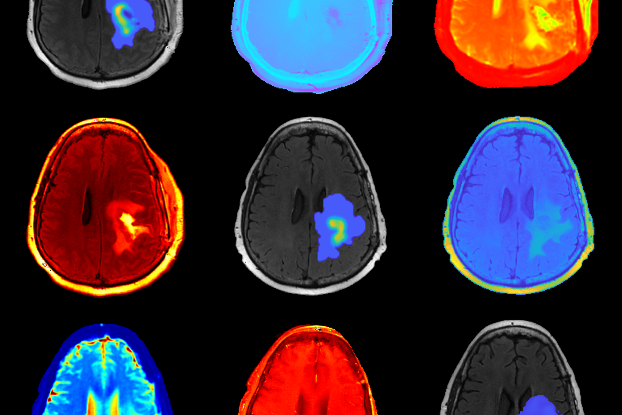
Issue 160: In our recent efforts to translate our pre-clinical models of glioma growth and response to the clinical setting we employed multiparametric MRI collected before and after radiation therapy to initialize and parameterize our model of tumor growth and response. Anatomical imaging was used to estimate the tumor burden, while diffusion weighted imaging was applied to estimate cell density. We then applied our modeling framework to forecast response to chemoradiation for each patient and overlaid the predicted distribution of tumor cells on anatomical images. This effort was a collaboration between the Oden Institute (Hormuth and Yankeelov) and the M.D. Anderson Cancer Center (Al Feghali, Elliott, and Chung). See the full publication in Sci Reports here.