
Click the image below to view the full size version of this
cover.
Created by: Ryan J Murphy
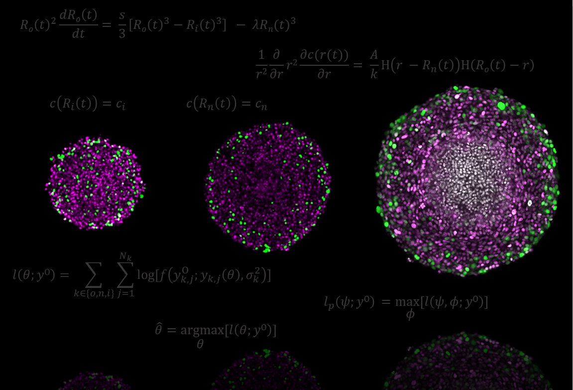
Issue 197: This artwork covers our recently published work in Communications Biology entitled Designing and interpreting 4D tumour spheroid experiments. The three experimental images of spheroids equatorial planes are representative of the three key phases of avascular tumour spheroid growth: phase (i) all cells able to proliferate; phase (ii) an outer region of proliferative cells and an inner region of proliferation-inhibited cells; and phase (iii) an outer region of proliferative cells, an intermediate region of proliferation-inhibited cells, and a necrotic core. Colours in the experimental images represent cell cycle status: G1 (magenta) and S/G2/M phases (green); and cell death (grey). To interpret these tumour spheroid experiments we measure outer, inhibited, and necrotic radii and use the seminal Greenspan mathematical model and statistical identifiability analysis (key equations shown in grey). Our approach allows us to determine maximum likelihood estimates and approximate 95% confidence intervals for parameters of Greenspan’s model across a range of experimental designs. We then identify experimental design choices that lead to reliable biological insights. Results are presented for human melanoma cell lines and our framework can be generalised to spheroids grown with different cell types and in different conditions.
© 2026 - The Mathematical Oncology Blog