
Click the image below to view the full size version of this
cover.
Created by: Jamie Auxillos, Stine Falsig Pedersen, Albin Sandelin, Rodolphe Marie, Yanfang Li, Roxane Crouïgneau, Arnaud Stigliani, and Yifan Dai
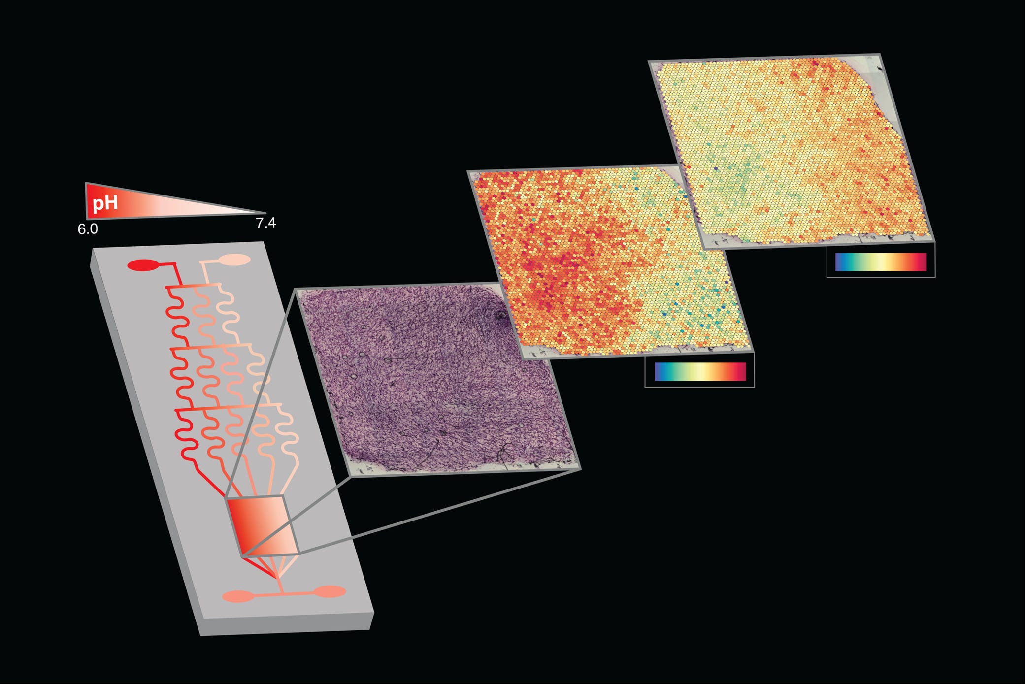
Issue 297: Despite the physiological and pathophysiological significance of microenvironmental gradients in cancer, tools for generating such gradients and analyzing their impact are lacking. Here, we present an integrated microfluidic-based workflow that mimics extracellular pH gradients characteristic of solid tumors while enabling high-resolution live cell imaging, and preserving the capacity to capture the spatial transcriptome. Our microfluidic device generates a pH gradient that can be rapidly controlled to mimic spatiotemporal microenvironmental changes over cancer cells embedded in a 3D matrix. The device can be re-opened allowing immunofluorescence analysis, as well as the transfer of cells and matrix to a Visium slide for spatially resolved analysis of transcriptional changes across the pH gradient. Using this method, we are able to identify the expression of genes such as ACTB (middle heat map), and FTL (top right heat map) from cells (in purple) grown and subjected to a pH gradient (from dark red corresponding to pH 6 to light pink for pH 7.4) in the microfluidics device. The ACTB gene, coding for the ubiquitous β-actin protein, shows a clear correlation between high expression and low pH while FTL, coding for the light chain of ferritin, has the opposite pattern (low expression with low pH). Check out the twitter thread on the paper.
© 2026 - The Mathematical Oncology Blog