
Click the image below to view the full size version of this
cover.
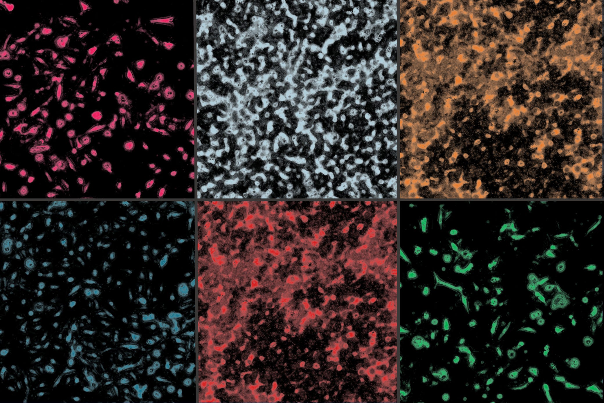
Created by: Maximilian Strobl
Issue 296: This week, I thought I would do a little behind the scenes about what I enjoy doing outside of the newsletter. In my work, I think a lot about how to calibrate and test mathematical models of cancer treatment scheduling. To truly understand the dynamical processes driving tumor cell kinetics, we need measurements at multiple time points. But these can be hard to come by in practice. This is because measurement can be a destructive process (e.g. tissue staining) and/or because if we want measurements every 4h that means someone has to collect these every 4h. Automated, time-series microscopy, in which a microscope is integrated with an incubator, so that the cells can be cultured and imaged automatically, is a simple yet powerful technique to get high resolution spatio-temporal data – and art! Shown here is a collage of bright field images I have been analyzing in recent weeks. More specifically, these are ovarian cancer cells treated with carboplatin and paclitaxel chemotherapy, and segmented with ilastik. For recent other examples of how to use such data to drive mathematical models, check out issues 290, 239, or 209.
© 2026 - The Mathematical Oncology Blog